
Iron instead of gadolinium: MRI contrast agent reveals heart scarring – and slows it
A Spanish team has developed an iron-based MRI contrast agent that makes fibrosis in heart tissue visible and slows its progression in a lab model. It has not yet been tested in patients.
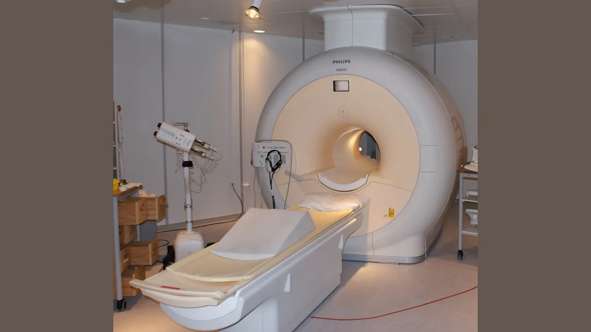
Symbolic image · Photo: Jan Ainali / Wikimedia Commons, CC BY 3.0 (cropped and resized)
In cardiac fibrosis, healthy heart tissue is progressively replaced by stiff connective tissue. Researchers at the CIC biomaGUNE research centre in San Sebastián and the Institute of Biomedicine and Biotechnology of Cantabria (IBBTEC) have developed a contrast agent for magnetic resonance imaging (MRI) that is designed both to reveal such changes and to treat them. The study was published in the journal Advanced Functional Materials (DOI 10.1002/adfm.78994).
Instead of gadolinium, which is used in almost all contrast-enhanced MRI scans, the team relies on iron: clusters of iron about 1.4 nanometres in size are encapsulated in a custom-designed protein. In 3D heart tissue grown from patients' heart cells, the agent accumulated in the scarred tissue, and its signal grew stronger as fibrosis increased. At the same time it reduced the production and deposition of collagen and prevented the tissue from stiffening. Its MRI contrast was "comparable to that of gadolinium-based agents", says lead author Lydia Martínez Parra.
So far it has only been tested on lab tissue models, not in animals or patients. Next, the researchers want to study how the agent is distributed and eliminated in the body and how it performs on a clinical scanner. There is "still a way to go to reach the patient", stresses the team led by Aitziber L. Cortajarena, scientific director of CIC biomaGUNE.