
PET in one to two minutes: total-body scanner delivers good image quality in study
A PET/CT scanner with a 128-centimetre field of view produced images after just one minute that experts rated higher than those of a conventional scanner, a Melbourne study found.
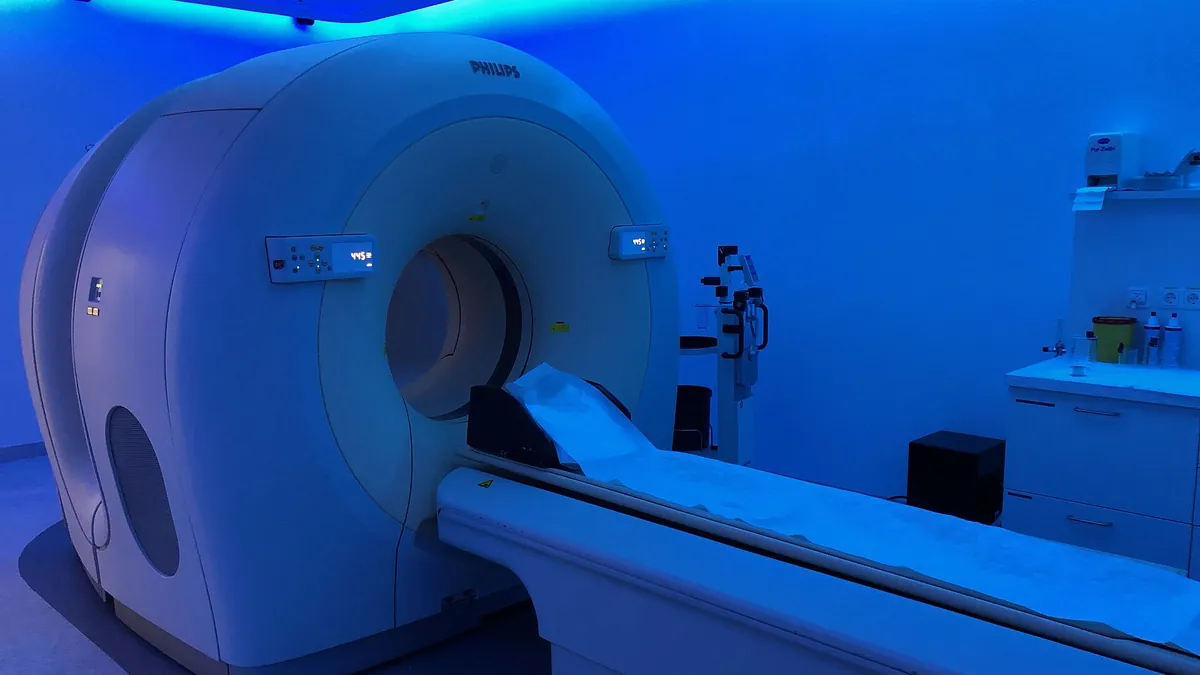
Symbolic image · Photo: Partynia / Wikimedia Commons, CC BY-SA 4.0 (cropped and resized)
A next-generation total-body PET scanner could significantly shorten examinations. That is the finding of the EQUIP study at the Peter MacCallum Cancer Centre in Melbourne, presented on 18 October at the congress of the European Association of Nuclear Medicine (EANM'26) in Vienna. It examined a GE HealthCare Omni PET/CT scanner with a 128-centimetre axial field of view compared with a conventional scanner.
24 patients were first scanned conventionally and then for ten minutes on the new device; images for 5, 3, 2 and 1 minute were also reconstructed from the data. Two nuclear medicine specialists rated the images on a five-point scale without knowing the acquisition time. Diagnostic quality averaged 5.0 points at 10 and 5 minutes, 4.9 at 3 and 2 minutes and 4.5 at one minute – the conventional scanner scored 3.9.
Even when acquisition time was cut by 90%, image quality exceeded the current standard of care, said lead author Cameron Pain, a nuclear medicine physicist at the Peter MacCallum Cancer Centre. Shorter scans could ease the burden on patients, reduce motion blur or allow lower doses of the radioactive tracer. A caveat: image quality was rated, not diagnostic accuracy; larger studies must show that small findings are not missed.